f WeR-3D Weight-Bearing CT | Clinical Diagnostics

WeR-3D Weight-Bearing CT

Redefining orthopaedic diagnostic standards through advanced natural weight-bearing imaging technology.

Why Weight-Bearing?

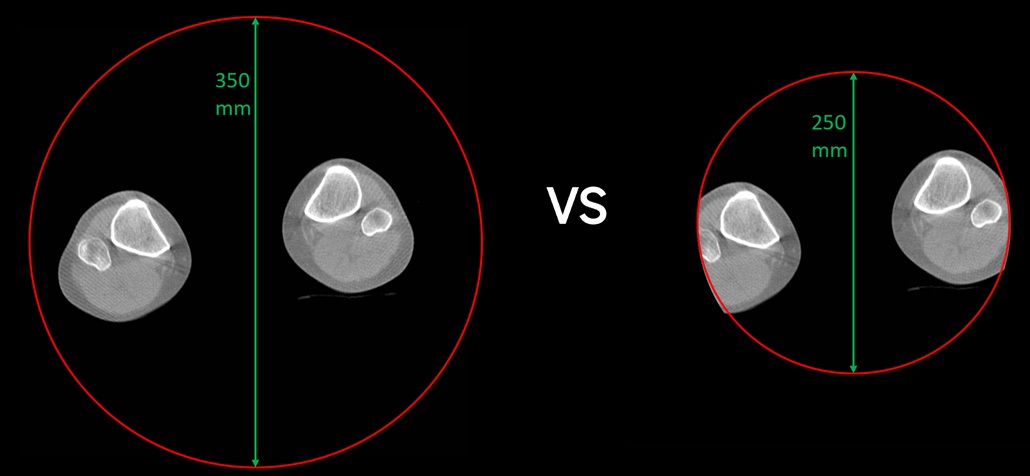
Traditional supine CT scans often fail to replicate true physiological stress on joints and the spine. This can lead to misdiagnosis, as critical alignment issues and joint space narrowing are only accurately visualized under natural, weight-bearing conditions.

Innovative Imaging Solutions

[产品图1]
[产品图2]

Radiation Safety Commitment

100%

Standard CT

85% Lower!
15%

WeR-3D

📚 Peer-Reviewed Evidence

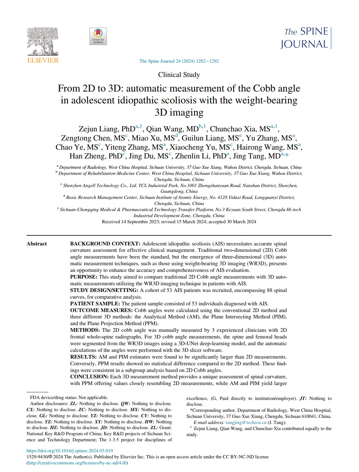
"From 2D to 3D: automatic measurement of the Cobb angle in adolescent idiopathic scoliosis with the weight-bearing 3D imaging."
Zejun Liang, Qian Wang, et al. | The Spine Journal (2024), 24(10), 1282–1292.

Clinical Imaging Comparison: Supine vs. Weight-Bearing

Case Study A

[平卧位影像1] [立位影像1]

Case Study B

[平卧位影像2] [立位影像2]

Why Angell 350mm FOV Matters?

FOV Comparison

Clinical Advantages

  • One-Stop Diagnosis: Captures entire joints in a single standing scan, making the process faster and reducing physical burden.
  • Comprehensive Visualization: Eliminates image truncation, ensuring a complete clinical picture and reducing the risk of misdiagnosis.
  • Enhanced Comfort: Streamlined workflow and natural standing posture provide a significantly more comfortable examination experience.
Full Spine

Full Spine

Hip Joint

Hip Joint

Lower Limbs

Lower Limbs

Bilateral Knees

Bilateral Knees

Bilateral Ankles

Bilateral Ankles

Clinical Research & Publications

学术论文 1

Research Study I

学术论文 2

Research Study II

学术论文 3

Research Study III

学术论文 4

Research Study IV

Inquire About WeR-3D

Prefer direct chat?

Chat on WhatsApp